- Summits
- Sponsorship
- Retreats
- Media Opportunities
- News
- About
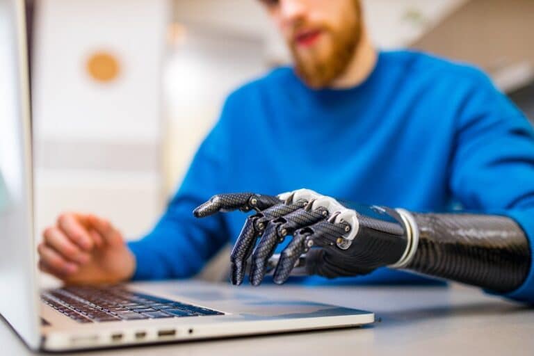
Researchers at the Massachusetts Institute of Technology (MIT) have developed a minimally invasive technology to allow users to control their bionic medical prostheses more accurately. Known as magnetomicrometry, the technology holds promise for people with muscle and bone pathologies, amputations, and spinal cord injuries.
While the technology already sounds exciting, it’s only been tested in-vitro (in controlled conditions in a lab) and in non-human subjects. After months of fine-tuning the technology, the researchers implanted the beads into turkey legs. They then had the turkeys run around an obstacle course – a real-world challenge. The results were highly encouraging; the system effectively measured and assessed muscle length.
In a published study in the journal Frontiers in Bioengineering and Biotechnology, the researchers documented how the magnetomicrometry technique fared against traditional techniques like fluoromicrometry in the real world. Fluoromicrometry (FM) enables high-bandwidth, high-precision (<90 μm) tracking of muscle fibres and has been used by researchers for over a decade. Detecting a high degree of correlation with fluoromicrometry, researchers were able to show that the technology was suitable for real-world use. Interestingly, the paper also shows that the technology may be more suitable for research, as FM requires specific laboratory constraints.
Long-term utility, another important concern with implants, seems to have been solved by the researchers. When investigating the effects of the magnetic beads on the body, the researchers found no adverse reactions to them in-vivo. This suggests the possibility of long-term use of the software tools.
Among the next few steps before these beads can enter the market are clinical trials. Tests in human subjects have yet to be underway. Still, noting that the technology has already been shown to be safe in vivo and the demonstrable utility of this in clinical scenarios, we may expect them soon.
One of the immediate opportunities that magnetomicrometry, or MM, offers is helping patients with amputated limbs, muscle weakness, and nerve damage recover natural motion post-injury. Signals from the magnetic beads can be sent to exoskeletal limbs and prostheses, enabling precise movements in response to isotonic contractions. Noting the absence of significant adverse reactions in previous tests, it offers the opportunity for long-term use, which will help drive costs down and facilitate practical usability.
MM also holds significant promise in research in muscle physiology and related fields. The researchers note that current technologies such as FM “require substantial post-processing time, hindering their use in longitudinal studies.” MM enables real-time data collection, allowing researchers to measure the behaviour of muscles in non-controlled environments more dynamically. Whether it’s by investigating the mechanisms of muscle activity or monitoring the degradation of muscle integrity in specific diseases, MM can facilitate significant progress in the way research on muscles is conducted.
Wearable sensors have become an integral part of life in 2022 as humans interact with more and more forms of technology in their daily routines. Researchers CR Taylor, SS Srinivasan, and others at the MIT Centre for Extreme Bionics, therefore, came up with an innovative idea to help amputees and other users of prosthetic limb attachments to carry out more natural movements with their prosthetics.
In a paper published in the leading science journal Science Robotics in August 2021, the pioneers proposed a new solution that could allow users to “continuously monitor muscle, tendon, and bone motion, allowing us to monitor performance, deliver targeted rehabilitation, and provide intuitive, reflexive control over prostheses and exoskeletons.” The most advanced prostheses at the time of their paper used a technology known as surface electromyography, where the electrical activity of muscle fibres is measured to provide a sense of the strength and degree of contraction of muscles. However, this technique fails to meet the level of accuracy needed to facilitate natural movements with prostheses or robotic limbs.
Their new technology, however, proposed a new form of sensors that could not just detect electrical activity, but also geometry and forces in a more accurate way. Magnetomicrometry, or MM, has now been tested in turkey models, with plans by the researchers to transition to human trials once the technology was shown to be effective and safe in-vivo. Positive results from these preliminary tests show promise for their new technology to become mainstream as the medical and tech sectors find more use cases for it.
Magnetomicrometry works by using magnets to make accurate measurements of muscles and dimensions in space. The technology involves the implantation of magnetic beads in muscles. The changing distance between the magnets in contractions and relaxations allows the system to measure movement accurately. The signals are then transmitted to a bionic limb or prosthesis, which then generates movement based on the information. When compared with current systems, magnetomicrometry offers significant advantages in that it can measure real movement instead of inferring movement from electrical activity.
In developing the technology, the researchers mounted these magnetic beads within the muscle. The beads move together as the muscle contracts and away from each other as the muscle relaxes. By generating a magnetic signal, the beads empower the robotic prostheses to mimic the user’s intended action more reliably.
Accuracy is also an important concern in medtech, and the device showed great promise in its accuracy in measuring muscle activity. By interacting directly with muscles geometrically, magnetomicrometry can track the nuanced movements of muscles with remarkable accuracy, allowing it to translate minute contractions into realistic movements. Magnetomicrometry allows users to implant multiple pairs of these beads into their muscles, enabling precise motion to be generated. Compared with traditional mechanisms, the technology did favourably well, implying that it could be a great alternative to existing solutions.
Magnetomicrometry also provides feedback relatively fast, with the researchers’ test recording a time of less than a millisecond for the tool to detect muscle length changes. With these speeds, there is the potential for real-time tracking of muscle movements and a more natural interaction of users of this technology with their environment.
That’s a wrap on 2022, and what a terrific year it has been! We at SiGMA Group are incredibly grateful to all the delegates, collaborators, sponsors and of course, the core team itself whose hard work and support is crucial to hosting the landmark quality events everyone has come to associate with the name SiGMA.
That being said, the world keeps on turning, and so shall the wheels of this event-hosting machine that was kickstarted in 2014. Coming right up is SiGMA Group’s premier event in Africa, with Nairobi 2023 set for next January. Visit our webpage for more information!